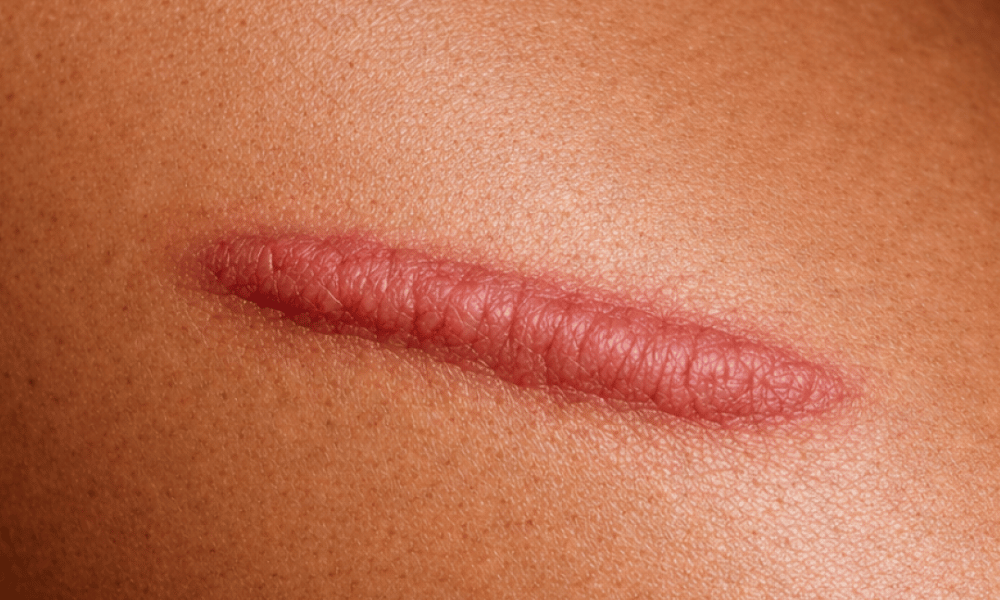
Surgery can seem like the most obvious treatment for a keloid. The reasoning is straightforward: the keloid is unwanted tissue, so remove it.
The difficulty is that a keloid is not simply a lump sitting on otherwise normal skin. It is the visible result of a healing response that behaves abnormally. Removing it creates a fresh wound in exactly the environment that produced the problem in the first place.
That does not mean surgery has no place. It means the question is rarely whether a keloid can be removed. It is whether removing it is the right step for this keloid, in this person, at this time — and what else needs to happen alongside it.
Can keloids be surgically removed?
Technically yes, and the operation itself is usually straightforward. The difficulty is what happens afterwards.
Excision on its own, with no additional treatment, carries a reported recurrence rate of forty-five to one hundred per cent, depending on the study, the site and the population [1]. That wide range reflects genuine variation between keloids and between patients. At the upper end, it means that in some settings essentially every keloid treated by surgery alone came back.
When a keloid does come back, it may be larger than the one removed.
This is the most important thing to understand about keloid surgery. Excision alone is generally not considered sufficient treatment. It is one part of a plan, and used on its own it carries a substantial risk of recurrence and of a larger scar.
Why does keloid surgery fail?
Keloids are increasingly understood as a long-term inflammatory disorder of the deeper layer of the skin, rather than simply an overgrowth of scar tissue [2].
In keloid-prone tissue, the inflammatory stage of healing does not settle on schedule. Inflammatory signalling continues, fibroblasts stay switched on, and collagen production carries on well past the point where it should have slowed. Several local factors keep that going: tension across the wound, slow healing, infection, foreign material, and the person’s own immune and genetic make-up.
Surgery removes the product of that process without changing the process itself. What remains is a new wound, under fresh tension, in tissue that has already shown a tendency to overproduce collagen. Recurrence in those circumstances is a significant concern.
There is a second consideration that is easy to overlook: how active the keloid is.
- An active keloid is growing, red, symptomatic and inflamed at its edges, where the growth is happening.
- A mature keloid is stable in size, paler, firmer and often no longer symptomatic.
Operating on actively inflamed tissue means creating a wound in an environment already primed to overproduce collagen. Assessing activity is therefore one of the first things done at consultation, and it affects both the timing and the content of the plan.
When is surgery appropriate?
Surgery may be considered in several situations, always as part of a wider plan.
Where the keloid is too large for other treatments to reduce. Topical medication, injections, laser and compression all work better on smaller lesions. Beyond a certain bulk, reducing the size surgically becomes the practical route to a scar that can then be controlled.
Where the keloid restricts function. A keloid crossing a joint or interfering with movement is a different proposition from one causing concern about appearance alone.
Where symptoms are severe and have not responded. Pain, intense itch and repeated ulceration within a keloid can be genuinely disabling, and are among the strongest predictors of reduced quality of life in people living with keloids [3].
Where the keloid sits on a narrow stalk. Earlobe keloids often do. This allows removal with a relatively short resulting wound and a correspondingly better outlook, particularly when combined with adjuvant therapies afterwards.
Where non-surgical treatment has been properly tried and has not achieved enough. Surgery is generally not the first step, and a keloid that has never had a proper course of conservative treatment has not yet shown that it needs an operation.
What determines whether it is a good option?
Several factors are weighed together, and no single one decides it.
Site. Keloids on the chest, shoulders and upper back sit in high-tension areas and carry a higher recurrence risk. Earlobes are low-tension and respond comparatively well.
Size and shape. A discrete, well-defined keloid is a more favourable prospect than a broad, diffuse plaque with unclear edges.
Activity. As above. Settling an actively inflamed keloid before operating changes the odds.
Previous treatment. A keloid that has already recurred after surgery behaves differently from one never operated on, and the plan has to account for what has already been tried.
Individual risk profile. Family history, age, keloids elsewhere, and any active inflammatory or systemic condition all contribute. Keloids arise most often between the ages of ten and thirty, and hormones appear to play a part, with keloids often becoming more active during puberty and pregnancy [4].
What can realistically be committed to. This is sometimes overlooked but matters. Keloid management after surgery runs for months to years, involving further treatment and follow-up. Someone who cannot commit to that is not well served by an operation.
What has to happen alongside surgery
The available evidence supports the principle that surgery combined with additional treatment produces substantially better outcomes than surgery alone.
Radiotherapy. Low-dose radiotherapy given shortly after excision is an established option. It works by suppressing the cells responsible for regrowth. Pooled recurrence after excision with radiotherapy falls to around thirteen and a half per cent, compared with the forty-five to one hundred per cent seen with surgery alone [5]. It is reserved for selected higher-risk cases, and the potential benefits are weighed carefully against the risks in each situation.
Injections into the scar. Intralesional infiltration of corticosteroid, sometimes combined with other agents, reduces inflammation and slows collagen production. Given before surgery it can reduce bulk and settle activity. Given afterwards it helps suppress regrowth during the vulnerable early months.
Compression. Sustained pressure after surgery is particularly useful for ear keloids. Custom-made pressure clips worn consistently after excision have been reported to prevent recurrence in around seventy per cent of patients [6], and excision combined with compression earrings has been reported with a recurrence rate of around ten per cent [7]. Compression can also be applied at most other body sites.
Laser. Fractional laser can improve texture and pliability. It is also used to help deliver medication into the scar, reaching dense tissue more evenly than injection alone.
Tension management. Careful deep-layer closure, avoiding closing under tension, and taping or silicone afterwards all reduce the mechanical stimulus that drives recurrence.
Which combination is appropriate depends on the individual scar, and is decided at consultation.
What the operation itself involves
The technical detail matters more here than in most scar surgery.
The keloid is removed, and the closure is designed to keep tension across the wound as low as possible. Sometimes that means orienting the closure along the skin’s natural tension lines. Sometimes it means rearranging local tissue to move tension away from the scar line. The deeper layers carry the load, so the skin edges sit together rather than being pulled together.
For larger keloids, some surgeons deliberately leave a rim of keloid tissue rather than cutting into normal skin, on the reasoning that the wound then sits within tissue that is already scarred. The evidence supporting this approach is limited, and whether it is appropriate is a matter of individual surgical judgement.
Additional treatment is planned before the operation, not decided afterwards. Where radiotherapy is part of the plan, its timing is coordinated with the surgery from the start.
What outcomes are realistic
The aim is a flatter, softer, less symptomatic scar, with a reduced likelihood of regrowth. It is improvement, not a return to unscarred skin.
Recurrence risk is reduced by combined treatment but not removed, and long-term follow-up is part of the treatment rather than an optional extra. Early regrowth is easier to control than established regrowth, which is one reason ongoing review matters.
Who should be doing this
Keloid surgery draws on several disciplines. It calls for surgical judgement about removal and closure, familiarity with injectable and laser-based treatments, and access to radiation oncology where radiotherapy forms part of the plan. It also calls for judgement about when surgery is not the right answer.
A recurring challenge in keloid management is that surgery performed on its own carries a high risk of recurrence. For this reason, surgery is often best considered as part of a broader treatment plan, with patients informed from the outset about the likelihood of recurrence and the role of additional treatment.
Scarless – The Clinic provides specialist keloid assessment and multimodal management, including surgery with planned adjuvant therapy, in Sydney. Get in touch to arrange a consultation if appropriate.
All procedures carry risks and potential complications. A comprehensive consultation and clinical assessment are required to determine suitability. Individual results may vary.
References
- Surgical excision with adjuvant therapies in the management of keloids: a systematic review. Medicina. 2026;62(5):916.
- Ogawa R. Keloid and hypertrophic scars are the result of chronic inflammation in the reticular dermis. Int J Mol Sci. 2017;18(3):606.
- Bijlard E, Kouwenberg CAE, Timman R, et al. Burden of keloid disease: a cross-sectional health-related quality of life assessment. Acta Derm Venereol. 2017;97(2):225–229.
- Limandjaja GC, Niessen FB, Scheper RJ, Gibbs S. The keloid disorder: heterogeneity, histopathology, mechanisms and models. Front Cell Dev Biol. 2020;8:360.
- Adjuvant radiotherapy following surgical excision of keloids: a systematic review of dose, fractionation, and recurrence. Life. 2026;16(5):770.
- Efficacy of custom-made pressure clips for ear keloid treatment after surgical excision. J Plast Reconstr Aesthet Surg. 2016;69(2):272–275.
- Compression therapy for keloid scars: a systematic review and meta-analysis. Plast Reconstr Surg Glob Open. 2024.